Antiviral Proteins
A major area of interest has been the discovery and characterization of proteins that comprise intrinsic cellular antiviral defenses. The lab revealed that non-human primate cells possess an activity that targets incoming lentiviral capsids whose antiviral activity varied in a species-dependent manner. The protein responsible for this activity was later identified by others as TRIM5, and the lab performed key studies mapping determinants of its specificity and providing key insights into the TRIM5 mechanism of action.
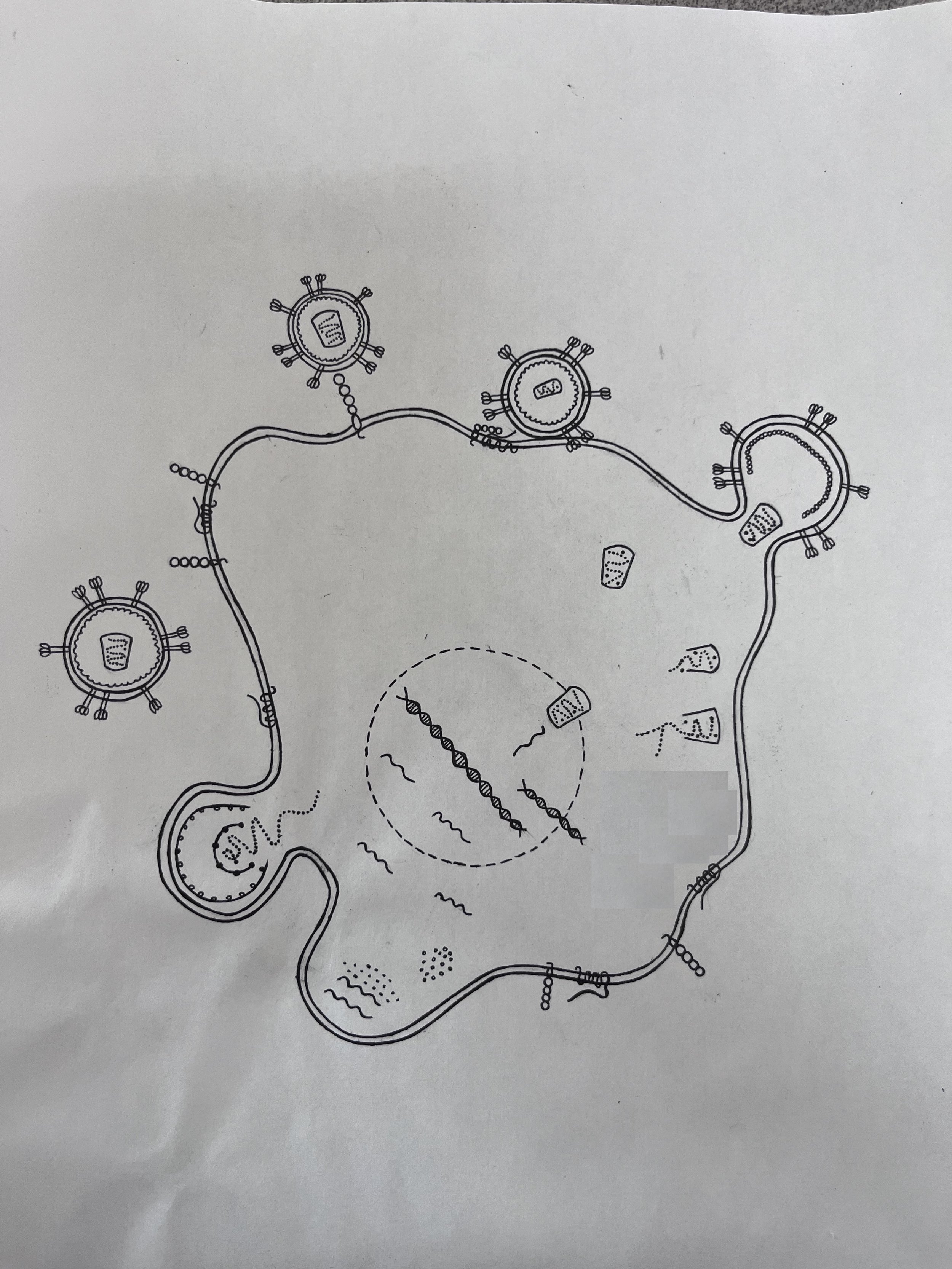
The group discovered a protein that traps a remarkably set diverse enveloped virus particles on the surface of cells, that they termed ‘Tetherin’. The lab showed how Tetherin inserts itself into the lipid envelope of virions to cause their entrapment, in a way that explains why the overall protein structure, and not the primary sequence, of Tetherin is important for activity. The lab showed how Tetherin evolved from a protein of unrelated function, that the HIV-1 accessory protein Vpu antagonized the action of Tetherin, and that simian immunodeficiency viruses (SIVs) lacking a Vpu protein often employ another viral accessory protein, Nef, as a Tetherin antagonist. The specificity of these viral antagonists is governed by host species-dependent Tetherin sequence variation. The team have also discovered a number of additional host proteins, including CNP, AhR, IDO1, TRIM56 and TRIM69, that either induce or have direct antiviral activity against HIV-1 or other viruses.
The lab found that RNA molecules that have too many CpG dinucleotides are recognized as non-self by cell intrinsic mechanisms and depleted from the cytoplasm. The recognition of CpG rich RNA’s as non-self was enabled by the progressive purging of CpG dinucleotides from the genomes of tetrapods that occurred over hundreds of millions of years. The lab identified zinc-finger antiviral protein (ZAP) as the protein responsible for the recognition of CpG rich foreign RNA, and showed that ZAP binds directly to CpG containing RNA to trigger its cytoplasmic depletion. With collaborators, the lab showed the structural basis for ZAP’s ability to discriminate of CpG-rich RNA, revealing that ZAP has a dinucleotide binding pocket that can accommodate a CpG, but no other dinucleotide. The team has also found that designing viral genomes with optimal numbers, context and spacing of CpG dinucleotides generates rationally and conditionally attenuated viruses that can work well as vaccines in model systems. Currently the lab is working to define the molecular details of how viral RNA is depleted following its recognition by ZAP.
Among the proteins discovered by the lab to exhibit antiviral activity is the interferon induced protein Mx2, which prevents capsid-dependent entry of HIV-1 preintegration complexes into the nucleus, The lab showed that Mx2 antiviral activity was dependent on nucleoporins - through a detailed examination of HIV-1 nuclear entry, the group showed that there are likely multiple independent paths by which HIV-1 can access the nucleus and that Mx2 inhibits subset of nuclear entry pathways that are preferentially exploited by HIV-1. Building on this work, the lab is developing new methods to track virus particles and subviral structures following viral entry and during nuclear import.
The lab continues to pursue the identification of host proteins with antiviral activity against HIV-1 and other viruses, and works to understand mechanistically how they inhibit viral replication. This work has recently extended into respiratory viruses in general and coronavirus in particular.
Animal Models
While species-specific barriers might protect humans from numerous viral assaults, they also pose a problem in developing animal models for human disease. Studies of how primate lentiviruses such as HIV-1 interact with cellular proteins has allowed the lab to develop an HIV-1-strain that, for the first time, is able to cause AIDS-like disease in a non-hominid, specifically a macaque species commonly used in laboratory research. This simian-tropic HIV-1 generates high levels of acute viremia, and progression to disease can be controlled by manipulating CD8+ cells. This approach enables the extremes of human disease outcomes (rapid progression and elite control) to be recapitulated in this animal model. This novel HIV-1 animal model has enabled the evaluation of HIV-1-specific therapeutics and prevention strategies including the demonstration that a new capsid inhibitor can provide effective protection from challenge. Currently, the lab is continuing to refine this model and is using it to determining the role of interferons in controlling HIV-1 replication in vivo.
The development of the HIV-1 macaque model and the description of how HIV-1 adapted to replicate in macaques, has illuminated how antiviral proteins are key in limiting virus host range. It has allowed the detailed characterization of such antiviral proteins in macaques and identification of novel variants amenable to structural studies. Indeed, with collaborators, the lab contributed to solving the first crystal structure of an antiviral cytidine deaminase (APOBEC3H) in a complex with an RNA target. The structure revealed a unique mode of protein-RNA interaction that drives the specific incorporation of APOBEC3H into viral particles.
The lab has also generated a number of recombinant vesicular stomatitis viruses with envelope proteins from other viruses including HIV-1 and coronaviruses. These are similar in design to recombinant virus vaccines, but can be used in immunodeficient mice to evaluate the effectiveness of vaccines in and therapeutic antibodies.
HIV-1 gene expression
The lab showed that that a chromosome periphery protein, CCDC137/cPERP-B, is targeted for depletion by the HIV-1 accessory protein Vpr. CCDC137 depletion causes G2/M cell-cycle arrest and enhances HIV-1 gene expression, particularly in macrophages. The lab is conducting a number of studies both in cell culture and in newly developed mouse model systems to understand how HIV-1 latency is regulated by cis-acting genomic contexts as well as trans-acting proteins using: 1) global synonymous mutagenesis to reveal the presence of several diffusely distributed and discrete RNA sequences that suppress the use of cryptic or canonical splice sites and 2) genetic screens to identify trans-acting proteins that regulate HIV-1 splicing and investigating the mechanisms by which candidate splicing regulators function.
Paleovirology
The lab pioneered the field of paleovirology, showing that an extinct retrovirus (HERV-K) could be ‘resurrected’ in functional form by reconstructing a pseudo-ancestral sequence, deduced from defective molecular ‘fossils’ that are present in modern human genomes. The lab also uncovered evidence of ancient interactions between APOBEC3 cytidine deaminases and extinct retroviruses in the form of hypermutated endogenous proviruses in humans and chimpanzees. The lab made the first identifications of entry receptors for extinct viruses using ‘resurrected’ viral envelope proteins, specifically chimpanzee endogenous retrovirus-2 and human endogenous retrovirus-T (HERV-T). The lab also showed that the human genome contains a defective HERV-T envelope gene whose product is capable of inhibiting infection mediated by ancient HERV-T envelope, that possibly contributed to HERV-T extinction.
Virus particle assembly and release
A seminal set of studies that included contributions from the lab elucidated the mechanisms by which so-called ‘late-budding’ domains enable the release of enveloped virus particles from infected cells. Specifically, the lab contributed to the discoveries of key roles for Tsg101, ALIX, HECT-ubiquitin ligases, ubiquitin and the ESCRT pathway in the budding and release of HIV-1, Ebola virus and other enveloped viruses. For a time, the subcellular location at which HIV-1 particle assembly occurs was controversial. The lab resolved this question definitively, demonstrating clearly that HIV-1 particles assemble at the plasma membrane. Building on that work, the lab collaborated to develop novel imaging techniques to observe and measure virion assembly. These techniques allowed the genesis of individual HIV-1 particles to be visualized in real time, in living cells - the first time this had been accomplished for any virus. This advance enabled unique studies of the assembly of individual HIV-1 virions allowing the visualization and quantification of the dynamics of viral genomic RNA movement and encapsidation, as well as the recruitment of ESCRT proteins to sites of virion release.
The lab also developed new biochemical and crosslinking-sequencing approaches to reveal, in unprecedented detail, how viral proteins and RNA interact during particle assembly. This work redefined how HIV-1 packages its genome, suggesting that the unusual nucleotide composition of the HIV-1 genome drives viral RNA interaction with assembling virion components. In recent work, the lab has shown that assembly of a nascent Gag lattice is required for recognition of the RNA packaging signal. These new approaches also uncovered a striking and specific interaction between the HIV-1 matrix domain of Gag and tRNA in the infected cell cytoplasm. With collaborators the lab solved a structure of an HIV-1 matrix:tRNA complex and showed that tRNA controls the subcellular localization of HIV-1 Gag protein, and thereby regulates particle assembly. The lab continues to develop new fluorescence microscopy approaches to study HIV-1 assembly.
Antibodies as drivers of viral glycoprotein evolution
During the COVID19 pandemic the lab created tools to understand the antibody response to the SARS-CoV-2 spike protein, viral spike evolution and genetic barriers to viral escape from antibodies. These studies revealed, in real time, how neutralizing antibodies varied and evolved after SARS-CoV-2 infection, administration of various vaccines, breakthrough infections and ever-increasing combinations thereof. The lab showed how these antibody responses are primarily responsible for driving spike evolution, recurrent waves of variants and how epistasis, as exemplified in the omicron lineage, lowers the genetic barrier to escape from neutralizing antibody. Current studies in this area are focused on generating sarbecovirus vaccines that elicit antibodies with the maximum possible breadth by asking whether it is possible to selective stimulate B-cells with broadly reactive B-cell receptors or alternatively whether broadly neutralizing polyclonal sera inevitably contain numerous distinct neutralizing specificities that act together to generate breadth. Additionally, the lab is conducting comparative studies with HIV-1 to understand the nature of the generic barrier to neutralizing antibody escape in the context of therapeutic antibody administration.
Coronavirus replication and host interactions
Coronaviruses have emerged as important real and potential human pathogens and identifying host cell factors important for coronavirus tropism and reproduction. The lab identified VPS29, a component of the retromer complex, as a required host protein for infection by HCoV-OC43, SARS-CoV-2, other endemic- and pandemic-threat coronaviruses, as well as ebolavirus. VPS29 was required for correct endosome morphology and acidity - its absence attenuated the activity of endosomal proteases, explaining its role the entry of some viruses. The lab showed that a SARS-CoV-2 accessory protein, ORF7a depletes MHC-I from the cell surface by preventing assembly of the MHC-I peptide loading complex and causing retention of MHC-I in the endoplasmic reticulum. In so doing, ORF7a inhibits antigen presentation. The lab is also developing a number of genetic systems and tagged coronaviruses for studying coronaviruses, and is deploying then to study various aspects of coronavirus cell and molecular biology.


